
Translate this page into:
Bilateral Milian’s Ear Sign: A Rare Presentation to Dermatologist
*Corresponding author: Samruddhi Naresh Chopkar, Department of Dermatology, Venereology and Leprosy, Indira Gandhi Government Medical College and Hospital, Nagpur, Maharashtra, India. samruddhichopkar159@gmail.com
-
Received: ,
Accepted: ,
How to cite this article: Chopkar SN, Ambade G, Rathi S, Mishra D. Bilateral Milian’s Ear Sign: A Rare Presentation to Dermatologist. Vidarbha J Intern Med. 2023;33:111-2. doi: 10.25259/VJIM_3_2024
Dear Editor,
Erysipelas is a cutaneous bacterial infection involving the upper dermis and/or superficial lymphatics.[1] Its diagnosis overlaps with cellulitis, which is the bacterial infection of the deeper dermis and subcutaneous fat, characterised by raised tender erythema. When the erythema spreads to involve the pinna, it is more likely to be erysipelas than cellulitis due to the absence of subcutaneous tissue in the pinna. This differentiating feature is known as ‘Milian’s ear sign’.[2] Bilateral ear erysipelas is rare.[3] Hereby, we report this case of bilateral Milian’s ear sign for its rarity and uncommon presentation to dermatologists.
A 47-year-old male patient presented to the dermatology department with complaints of fever with painful reddish swelling on both ears [Figure 1]. There was a history of head trauma six months back for which he was treated surgically. There was no complaint of any discharge from ear or hearing loss. Physical examination revealed tender oedematous, erythema with a few vesicles on bilateral ear pinna [Figure 2a and b]. There were no abnormalities of the external auditory canal and on otoscopic evaluation. ‘Milian’s ear sign’ was positive bilaterally [Figure 1]. The haemogram showed leucocytosis. Tzanck smear showed neutrophils with no multinucleated giant cells [Figure 3]. On the basis of the clinical examination, erysipelas and relapsing polychondritis were kept as the closest differential diagnosis. The patient was treated with oral amoxicillin and clavulanic acid combination at a dose of 625 mg twice daily for 7 days and his symptoms fully resolved over a duration of 7 days. Finally, a clinical diagnosis of bilateral ear erysipelas was made.
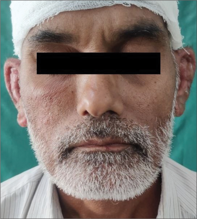
- A 47-year-old male patient presented with painful reddish swelling of both ears.
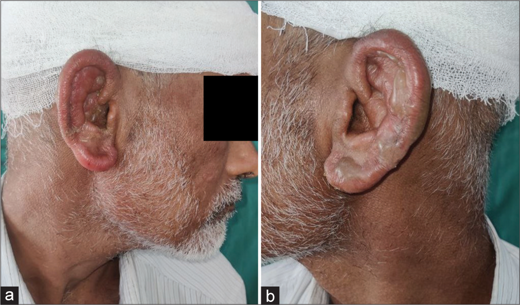
- (a and b) Clinical examination showing diffuse tender oedematous, erythema with a few vesicles on bilateral ear pinna (Milian’s ear sign positive) of the right and left ear, respectively.
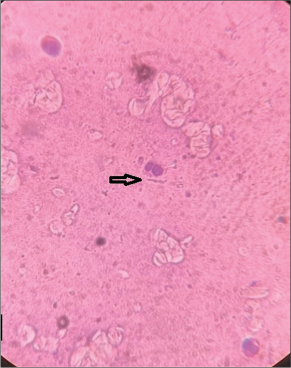
- Tzanck smear showed neutrophils with no multinucleated giant cells (on 10× microscopy with black arrow).
Erysipelas is a common bacterial infection caused by group A beta-haemolytic streptococcus, Streptococcus pyogenes A, of the dermis which may involve superficial lymphatics.[4] It is characterised by a well-demarcated, tender and raised area of erythema. ‘Milian’s ear sign’ is a sign of otic involvement of infection, differentiating erysipelas from cellulitis.[2] The definitive treatment for this infection is oral/injectable penicillin or clindamycin.[5]
Bilateral ear erysipelas is rare and its presentation to the dermatologist is also rare, but it should be considered in the differential diagnosis of patients with red, painful ears.
Ethical approval
The Institutional Review Board approval is not required.
Declaration of patient consent
The authors certify that they have obtained all appropriate patient consent.
Conflicts of interest
There are no conflicts of interest.
Use of artificial intelligence (AI)-assisted technology for manuscript preparation
The authors confirm that there was no use of artificial intelligence (AI)-assisted technology for assisting in the writing or editing of the manuscript and no images were manipulated using AI.
Financial support and sponsorship
Nil.
References
- Erysipelas: Recognition and Management. Am J Clin Dermatol. 2003;4:157-63.
- [CrossRef] [PubMed] [Google Scholar]
- Eponymous Signs in Dermatology. Indian Dermatol Online J. 2012;3:159-65.
- [CrossRef] [PubMed] [Google Scholar]
- Bilateral Milian's Ear Sign of Erysipelas. Intern Med. 2017;56:2381-2.
- [CrossRef] [PubMed] [Google Scholar]
- Streptococcal Cause of Erysipelas and Cellulitis in Adults. A Microbiologic Study Using a Direct Immunofluorescence Technique. Arch Dermatol. 1989;125:779-82.
- [CrossRef] [PubMed] [Google Scholar]


